
What is palmoplantar pustulosis?
‘Palmoplantar’ literally refers to the palms of the hands and soles of the feet. One of the characteristics of palmoplantar pustulosis is the formation of ‘pustules’ (red area containing pus) on the palms of the hands and soles of the feet (figure 1). The pustule formation continuously takes place under the cycle that usually starts as tiny blisters on the aforementioned area which gradually becomes filled with pus inside and finally sheds. The affected skin usually shows reddish change (Photo 1). Both palms of the hands and soles of the feet can be involved; however, it can affect any combination of the palms or feet.
The condition can also cause the fingernails and/or toenails to be deformed.
Having these eruptions of palmoplantar pustulosis can accompany pain and/or itchiness and can negatively affect the patient’s quality of life.
In the rare cases, the midline joint between the right and left clavicles (sterno-clavicular joint) and some other joint inflammation may accompany the skin symptoms, the condition known as palmoplantar osteo-arthritis.
The condition can also cause the fingernails and/or toenails to be deformed.
Having these eruptions of palmoplantar pustulosis can accompany pain and/or itchiness and can negatively affect the patient’s quality of life.
In the rare cases, the midline joint between the right and left clavicles (sterno-clavicular joint) and some other joint inflammation may accompany the skin symptoms, the condition known as palmoplantar osteo-arthritis.
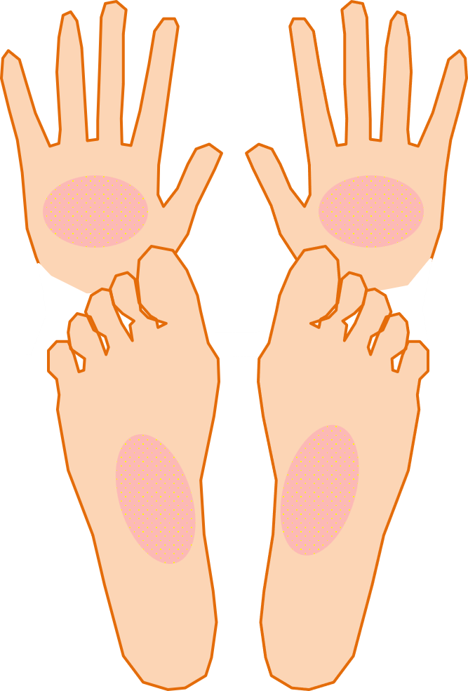
Figure 1. In palmoplantar pustulosis, patients show redness and pustule formation on the palms of the hands and soles of the feet.
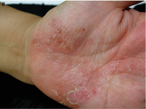
Photo 1. Murakami M et al. J Invest Dermatol 2010; 130: 2010-6
The diagnosis of palmoplantar pustulosis
The diagnosis of palmplantar pustulosis can be made by a dermatologist upon seeing if the affected area shows typical tiny blisters and pustules on the palms of the hands and the soles of the feet, however it is occasionally difficult to differentiate from other dermatological conditions such as dyshidrotic eczema (a type of hand eczema).
In order to make a correct diagnosis, we may propose a skin biopsy (to cut out the blister or pustule under local anesthesia). We consider the correct diagnosis as very important since this can be a first step to appropriate treatment.
In order to make a correct diagnosis, we may propose a skin biopsy (to cut out the blister or pustule under local anesthesia). We consider the correct diagnosis as very important since this can be a first step to appropriate treatment.
How many patients are suffering?
The prevalence of palmoplantar pustulosis seems to vary by region of the world, race, and gender.
Statistically it is speculated that we have 1 to 2 patients with palmoplantar pustulosis among 1,000 people in Japan (1), meanwhile the rate is reported to be 1 to 2 out of 10,000 people in the US and the UK (2)(3). Thus, the prevalence seems to be disproportionately affecting Asian populations including Japan as compared with Europe and North America.
Female gender tends to have a higher prevalence than males according to many reports (Male to female ratio is almost 1 to 2 in Japan). Peak prevalence age is reported to be between 30’s and 50’s. Also, patients tend to seek medical attention in summer rather than in winter
(the patient usually experiences worsening of the symptoms in hotter seasons)(1).
(1)Kubota K et al. BMJ Open 2015: 5: e006450
(2) Frysz M,et al: Br J Dermatol. 2024;191:529-538
(3) Ramcharran D,et al: Adv Ther. 2023;40:5090-5101
Statistically it is speculated that we have 1 to 2 patients with palmoplantar pustulosis among 1,000 people in Japan (1), meanwhile the rate is reported to be 1 to 2 out of 10,000 people in the US and the UK (2)(3). Thus, the prevalence seems to be disproportionately affecting Asian populations including Japan as compared with Europe and North America.
Female gender tends to have a higher prevalence than males according to many reports (Male to female ratio is almost 1 to 2 in Japan). Peak prevalence age is reported to be between 30’s and 50’s. Also, patients tend to seek medical attention in summer rather than in winter
(the patient usually experiences worsening of the symptoms in hotter seasons)(1).
(1)Kubota K et al. BMJ Open 2015: 5: e006450
(2) Frysz M,et al: Br J Dermatol. 2024;191:529-538
(3) Ramcharran D,et al: Adv Ther. 2023;40:5090-5101
What causes palmoplantar pustulosis?
The exact cause of PPP is still inconclusive.
But many exacerbating factors have been reported to date. At the same time, according to several reports, getting rid of these exacerbating factors can result in the alleviation or even permanent disappearance of the symptoms.
Many times these exacerbating factors are correlated with cigarette smoking, odontogenic focal infection, and tonsillar focal infection.
But many exacerbating factors have been reported to date. At the same time, according to several reports, getting rid of these exacerbating factors can result in the alleviation or even permanent disappearance of the symptoms.
Many times these exacerbating factors are correlated with cigarette smoking, odontogenic focal infection, and tonsillar focal infection.
A) Smoking Cigarettes
The smoking rate of the patients with palmoplantar pustulosis is reported to be as high as 70% to 90% 4)-5). Of course, this does not necessarily mean that everyone that smokes always have risk to develop palmoplantar pustulosis. By the way, the smoking rate of habitual smokers in Japan is reported to be 14.8 % in 2024 6). Also, studies have shown that quitting cigarette smoking had reduced the number of pustules of the hands and feet 7).
4) Akiyama T et al. J Dermatol, 22: 930, 1995
5) Erikson MO et al. Br J Dermatol 138: 390, 1998
6) Ministry of Health, Labor and Welfare website: 2024, National health insurance, nutritional assessment resutls
7) Michaelsson G et al. J Am Acad Dermatol, 54: 737, 2006
4) Akiyama T et al. J Dermatol, 22: 930, 1995
5) Erikson MO et al. Br J Dermatol 138: 390, 1998
6) Ministry of Health, Labor and Welfare website: 2024, National health insurance, nutritional assessment resutls
7) Michaelsson G et al. J Am Acad Dermatol, 54: 737, 2006
B) Odontogenic Focal Infection
Odontogenic Focal Infection refers to an infection around the teeth including periodontal disease (also known as gum disease in which a space between a tooth and gum becomes wide enough to gradually make the tooth loose), or apical periodontitis (an infection at the tip of a tooth root). There is a study that reported the complication rate of odontogenic focal infection to be more than 80 % of those diagnosed palmoplantar pustulosis patients 8). Also, dental treatment for these focal infection is reported to be effective to alleviate the symptoms of palmoplantar pustulosis in 60 % to 80 % of patients.
8) Kouno M et al: J Dermatol 44: 695-698, 2017
8) Kouno M et al: J Dermatol 44: 695-698, 2017
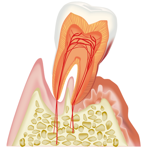
Image 2. In periodontal disease, the gum or jaw bones that support the teeth are gradually worn down due to chronic bacterial infection.
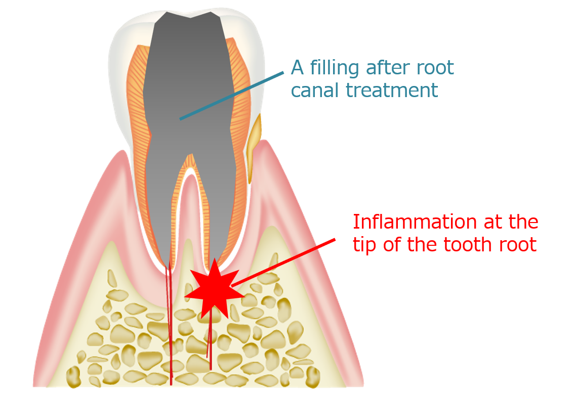
Image 3. Apical periodontitis is a condition in which an inflammation lesion develops in the root region of a tooth.
C) Tonsillar Focal Infection
In palmoplantar pustulosis, the tonsils can be a causative organ that may lead to the development and exacerbation of the skin symptoms on the palms and soles. Recurrent inflammation of the tonsils can cause the worsening of the symptoms, but the tonsils themselves can be involved in the symptoms of palms and soles regardless of the presence of inflammation. Also, some relief to complete disappearance of the skin eruptions of the palms and soles after surgical removal of the tonsils have been reported 9).
9) Kouno M,et al:J Dermatol 44:695-698,2017
9) Kouno M,et al:J Dermatol 44:695-698,2017
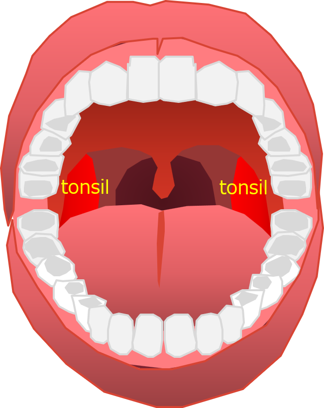
Image 4. The red structures on both sides at the back of the mouth are the tonsils.
Treatment of palmoplantar pustulosis
As for the treatment of palmoplantar pustulosis, we need to;
try to find and remove exacerbating factors in collaboration with other specialists such as an ENT doctor, and/ or a dentist.
2) topical or systemic treatment such as oral or injectable medications at a dermatology clinic.
At our clinic, both exacerbating factors and appropriate medications are simultaneously sought out for the symptom improvement.
try to find and remove exacerbating factors in collaboration with other specialists such as an ENT doctor, and/ or a dentist.
2) topical or systemic treatment such as oral or injectable medications at a dermatology clinic.
At our clinic, both exacerbating factors and appropriate medications are simultaneously sought out for the symptom improvement.
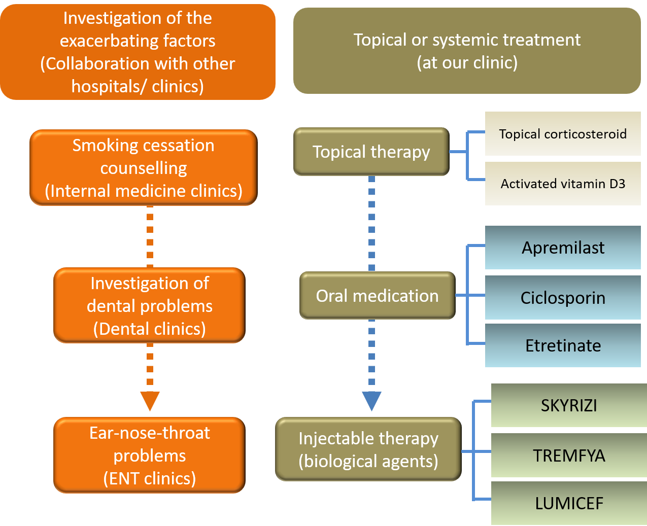
Figure 5. Overview of the Treatment Approacches for Palmoplantar Pustulosis
Investigation of the exacerbating factors of palmoplantar pustulosis
Trying to find and to treat the worsening factors of palmoplantar pustulosis plays a pivotal role as part of treatment in our approach against the disease since this can not only improve the symptoms but even lead to a cure. However, dealing with associated medical challenges such as quitting smoking, seeking dental treatment and the consideration of tonsil removal along with the collaboration with other medical specialists. At our clinic we proceed with such investigations with local credible internal medicine, dental and ENT clinics.
Smoking Cessation Counseling
The smoking rate of the patients with palmoplantar pustulosis is high and it is expected that kicking the habit of smoking can lead to the amelioration of the symptoms. If you have palmoplantar pustulosis and a smoking habit, we explain the importance of smoking cessation. If you feel difficulty stopping smoking, we might propose a reference to an affiliated nearby internal medicine clinic that provides smoking cessation counseling.
Investigation of dental problems
Treating gum disease or tooth root infection can lessen the symptoms or even cure the palmoplantar pustulosis. As for dental treatment, we propose to refer the patients to affiliated nearby dentists that pursue active treatment against correlated dental conditions.
Investigating ear-nose-throat problems
Sinusitis (an inflammation of the bone cavity around the nose) can be an exacerbating factor of palmoplantar pustulosis. Additionally, dental problems in the upper jaw bone can extend upwards to cause sinusitis. To reach the diagnosis of these problems, CT scan sometimes can be useful. If these conditions are suspected, we may propose you go to affiliated specialized diagnostic imaging clinic to have an imaging study.
薬剤による掌蹠膿疱症の治療
掌蹠膿疱症の薬剤による治療は、外用療法(塗り薬による治療)、内服療法(飲み薬による治療)、注射薬による治療(生物学的製剤による治療)があります。
外用療法(ぬり薬による治療)
掌蹠膿疱症に有効な外用剤には、ステロイド外用薬、活性型ビタミンD3外用薬の2種類があります。当院ではこれらを組み合わせたり、症状によっては単独で処方します。
内服療法(飲み薬による治療)
飲み薬による治療には、皮膚の角化異常を調節する作用のあるチガソン®(エトレチナート)があります。また、免疫調節作用をもち皮膚の細胞や免疫細胞の相互作用をおさえるオテズラ®(アプレミラスト)が2025年より新たに治療薬に加わりました。
注射療法(生物学的製剤による治療)
掌蹠膿疱症で使われる注射薬は、生物学的製剤とよばれ、現在3製剤が使用可能です。いずれもバイオテクノロジーにより作られた抗体製剤です。掌蹠膿疱症では、悪化因子の検索や除去、外用療法などで十分な効果が得られない患者さんに導入が検討されます。
掌蹠膿疱症では、IL-17(アイ・エル・17)、IL23(アイ・エル23)という細胞同士が情報を伝えるためのタンパク質が病態に関わっていると報告されています。生物学的製剤はこれらのいずれかのサイトカインをピンポイントで抑えることで効果を発揮します。
生物学的製剤の使用には、定期的な検査と、副作用に対応できる生物学的製剤承認の総合病院や呼吸器内科専門医による対応のとれる病院との連携が推奨されています。当院は生物学的製剤承認施設であり、これらを遵守し、診療にあたっています。
生物学的製剤の導入および維持投与においては、定期的な胸部X線撮影や胸部CT撮影、ウイルス性肝炎の有無調べる血液検査などが必要となります。胸部X線撮影や胸部CT検査は別途、当院と提携している他院で行っていただく必要があります。
生物学的製剤を用いた治療では、医療費が自己負担限度額を超える場合があります。医療費補助制度につき、正しい知識を持つことにより、医療費の負担を軽減し得る場合があります。詳しくは、医療費補助制度についての項目をご参照ください。
当院は、治療および医療費の補助制度につき正しい情報提供をし、安全な医療を行っていくことに努めています。
掌蹠膿疱症では、IL-17(アイ・エル・17)、IL23(アイ・エル23)という細胞同士が情報を伝えるためのタンパク質が病態に関わっていると報告されています。生物学的製剤はこれらのいずれかのサイトカインをピンポイントで抑えることで効果を発揮します。
生物学的製剤の使用には、定期的な検査と、副作用に対応できる生物学的製剤承認の総合病院や呼吸器内科専門医による対応のとれる病院との連携が推奨されています。当院は生物学的製剤承認施設であり、これらを遵守し、診療にあたっています。
生物学的製剤の導入および維持投与においては、定期的な胸部X線撮影や胸部CT撮影、ウイルス性肝炎の有無調べる血液検査などが必要となります。胸部X線撮影や胸部CT検査は別途、当院と提携している他院で行っていただく必要があります。
生物学的製剤を用いた治療では、医療費が自己負担限度額を超える場合があります。医療費補助制度につき、正しい知識を持つことにより、医療費の負担を軽減し得る場合があります。詳しくは、医療費補助制度についての項目をご参照ください。
当院は、治療および医療費の補助制度につき正しい情報提供をし、安全な医療を行っていくことに努めています。
表1. 掌蹠膿疱症で使用される生物学的製剤
掌蹠膿疱症の治療で使用される生物学的製剤を示します。赤字で示しているのが標的分子です。製剤により自己注射が可能な薬剤(家で自分で注射する薬剤)と、自己注射はできず、院内で医療スタッフにより注射を行う薬剤があります(表1: 表は右にスクロールできます)
|
製品名 (一般名)
|
標的分子 (さらに詳細な標的分子)
|
投与間隔*
|
自己注射
|
|
トレムフィア (グセルクマブ)
|
IL-23 (IL-23p19)
|
8週間
|
×
|
|
スキリージ (リサンキズマブ)
|
IL-23 (IL-23p19)
|
12週間
|
×
|
|
ルミセフ (ブロダルマブ)
|
IL-17 (IL-17RA)
|
2週間
|
〇
|
*各製剤は、導入初期にはローディングといって、薬剤の血中濃度を上げるため、表に記載されている投与間隔より短い間隔で注射スケジュールが規定されているものが多いです。表にある投与間隔は、ローディング後の投与間隔となります。